Our Products & Solutions
Gamma-ray imaging for security and nuclear sites, and dose tools for medicine — built on the same detector physics and Monte Carlo transport.
RadLens C1
A detector array, a depth camera and reconstruction software in one portable enclosure. Carry it in, switch it on, and watch the radiation appear in the room.
Product — first order secured from Incheon Main Customs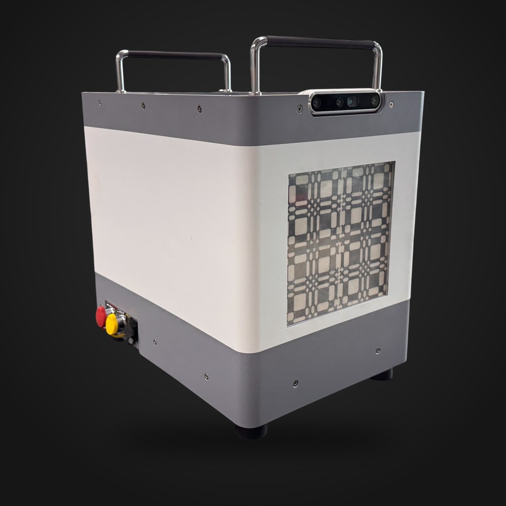
Capabilities
Live 3D radiation image
Drawn over the camera view with depth, so the signal sits on the object that carries it.
Sub-second acquisition
0.54 s ± 0.03 to a usable image. Short enough to sweep a space by walking through it.
Isotope identification
The energy spectrum resolves alongside the image, separating industrial sources from natural background.
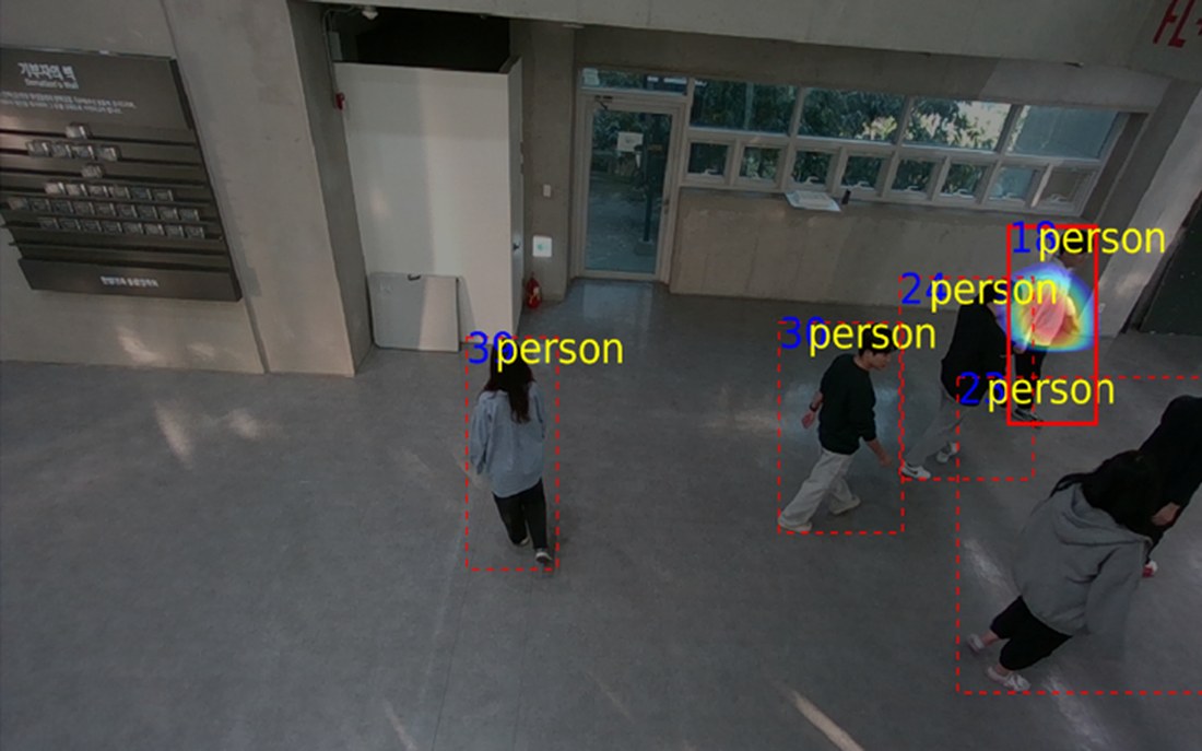
Detection and tracking
People and objects are followed frame to frame, so a source stays with the same carrier while it moves.
Sub-second imaging
Conventional gamma cameras wait and count. Our reconstruction produces a usable localisation from a much smaller event count.
| System | Imaging method | Acquisition time |
|---|---|---|
| RadLens C1 | Hybrid, with scene tracking | 0.54 s ± 0.03 |
| Commercial gamma camera A | Compton reconstruction | 83 s ± 50.6 |
| Commercial gamma camera B | Compton reconstruction | 81 s ± 33.5 |
Measured with a 137Cs point source, 10 µCi at 1 m (0.028 µSv/h), approx. 9° angular resolution. The RadLens C1 figure is measured in-house; comparison figures come from published literature and manufacturer datasheets, under the conditions stated there.
How it works
Coded aperture
A patterned mask casts a different shadow for each source direction. Fast and effective at lower photon energies.
Compton reconstruction
At higher energies, photons that scatter once and are absorbed again constrain the source to a cone; many cones meet at the source.
Scene registration
A co-axial depth camera supplies the room geometry, turning an angular distribution into a position on a real surface.
Where it is used
Customs and ports
Find which container, and where inside it, without unpacking the load.
Decommissioning
Map contamination over the real structure before people go in to cut it apart.
Reactors and SMRs
Locate leaks and hot spots during maintenance, from a safe standoff.
Emergency response
Resolve a reported source quickly, in a place nobody has mapped.
Specification
- Imaging method
- Hybrid gamma-ray imaging — coded aperture and Compton reconstruction
- Detector
- Scintillator array with position-sensitive readout
- Scene input
- RGB-D camera, co-registered with the radiation image
- Acquisition
- 0.54 s ± 0.03 (137Cs, 10 µCi at 1 m)
- Angular resolution
- approx. 9°
- Field of view
- ±90° horizontal, ±60° vertical
- Spectroscopy
- Energy spectrum with isotope identification and classification
- Scene processing
- Object detection and multi-object tracking
- Output
- 3D activity distribution over live video; source position, isotope list and track log
- Form factor
- Portable enclosure with carry handles; on-board processing; battery operation
- Status
- Evaluated on site; first order secured from Incheon Main Customs, for delivery in 2027
- Supply
- Instrument purchase, including maintenance; public-sector buyers through government procurement. Pilot evaluations on request.
Figures are measured in-house under the reference condition stated. Datasheets, acceptance test conditions and integration documentation are available on request.
Phantoms & IR Dose
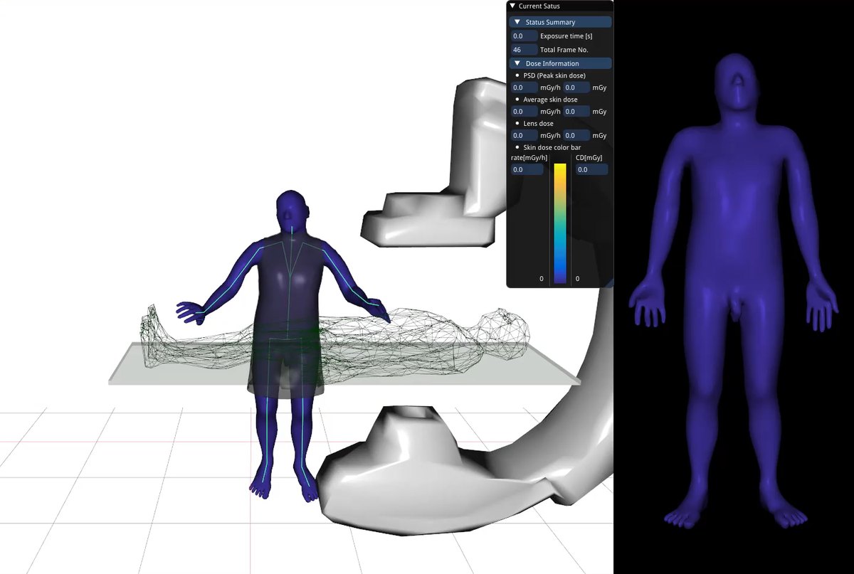
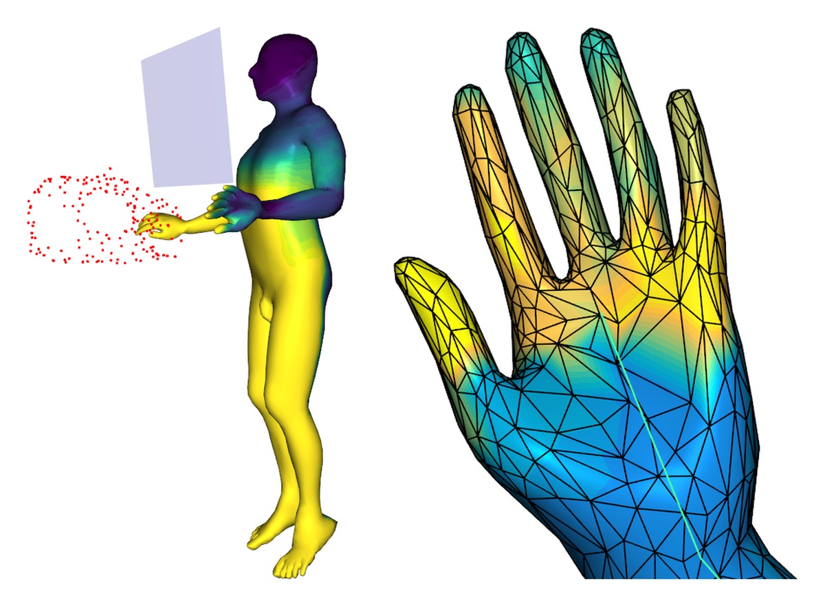
Our team developed the adult mesh-type reference phantoms that the International Commission on Radiological Protection adopted as ICRP Publication 145. We build on that work commercially in two ways: personalised phantoms, generated to an individual’s body size and posture, and a dose assessment program for interventional radiology that computes the dose to staff during a procedure.
Product — personalised phantoms and IR dose assessment software in commercial supply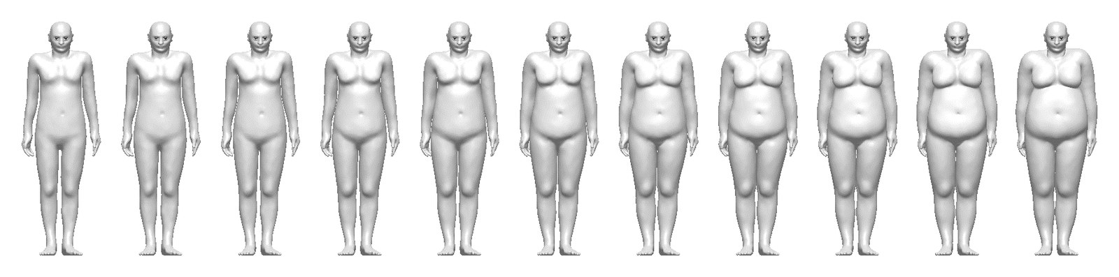
What we supply
Personalised phantoms
Mesh-type human models generated to a person’s height, weight and posture, with organ-level detail, for Monte Carlo dose calculation.
IR dose assessment program
Follows the operator beside the C-arm and computes peak skin dose, average skin dose and lens dose while the procedure runs.
Who uses it
Hospitals and IR suites
Staff dose per procedure, per person, rather than a single badge reading.
Medical physics and research
Organ dose studies across body sizes that a single reference body cannot represent.
Regulators
Dose assessment grounded in the international reference models.
Proton Dose Verification
A prompt gamma camera that measures where each spot of a proton beam actually stops, and software that turns those measurements into the dose that was really delivered and checks it against the treatment plan in real time. For particle therapy centres.
In clinical evaluation — phantom studies completed at the Samsung Medical Center proton therapy centre, and demonstrated to Sumitomo Heavy Industries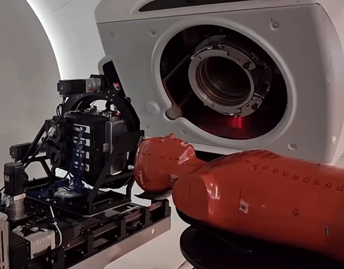
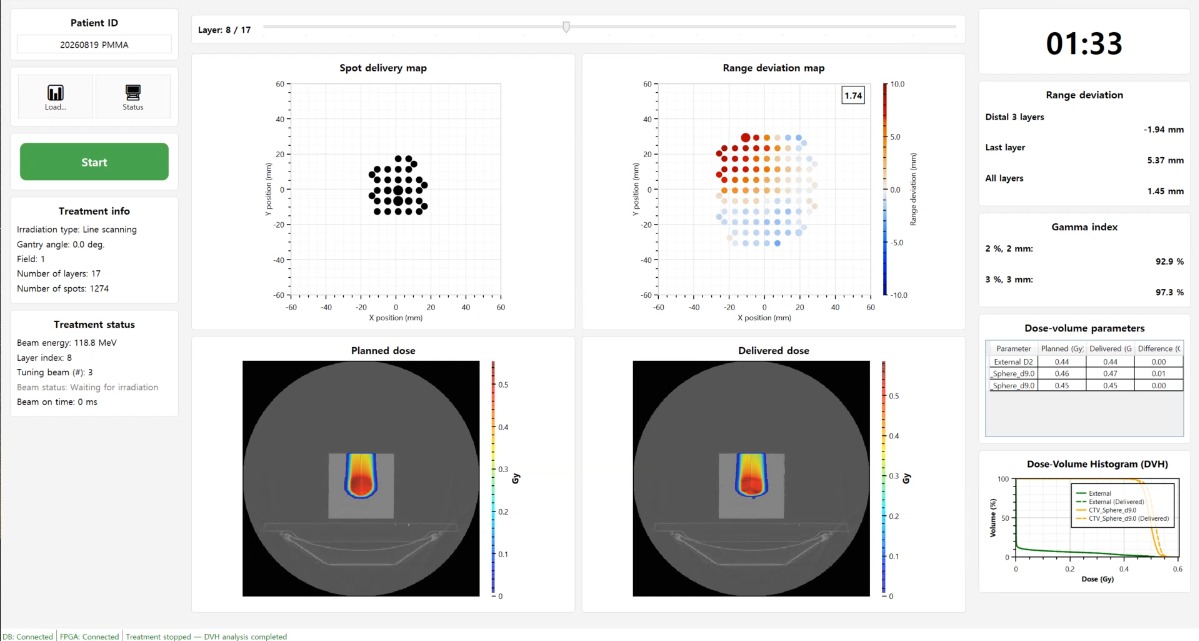
What the operator sees
Spot delivery map
Every delivered spot of the current energy layer.
Range deviation map
Measured minus planned range for each spot, in millimetres.
Planned vs delivered dose
The reconstructed delivered dose beside the plan, on the patient CT.
Gamma index and DVH
Agreement and target coverage, updated as the treatment runs.